PRESERFLO MicroShunt Revision: Managing a Fibrotic Bleb After Filtration Failure
How do you approach a failed PRESERFLO MicroShunt in an eye with extensive conjunctival scarring? This surgical case explores the revision of a PRESERFLO MicroShunt following progressive filtration failure associated with a flat fibrotic bleb and significant conjunctival fibrosis.
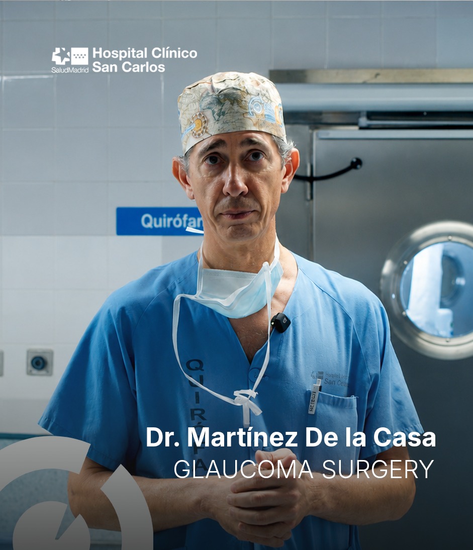
Access via the Ophthalmology Radar platform. Registration required.
About this surgical case
An 82-year-old patient undergoes revision of a PRESERFLO MicroShunt implanted two years earlier following progressive filtration failure associated with a flat fibrotic bleb and significant conjunctival fibrosis.
This case provides a detailed, step-by-step view of the surgical decision-making and technique involved in restoring effective filtration while preserving the existing MicroShunt — a clinically relevant scenario for any surgeon working with modern glaucoma drainage devices.
Surgical approach
The procedure demonstrates a structured approach to bleb revision in the context of advanced fibrosis:
Careful conjunctival dissection through scarred tissue
Release of the distal end of the MicroShunt from the fibrotic capsule
Confirmation of flow through the device
Mitomycin C application to reduce recurrent fibrosis
Fixation of the MicroShunt to the sclera
Layered closure of Tenon's capsule and conjunctiva
Tissue management is key to restoring filtration while preserving the existing MicroShunt.
What you will learn
Identify and manage a fibrotic bleb following PRESERFLO MicroShunt surgery.
Understand surgical bleb revision and subconjunctival fibrosis release.
Restore effective filtration while preserving the existing MicroShunt.
Recognize critical surgical steps to reduce the risk of recurrent bleb failure.
Skills you will develop
Bleb assessment, fibrosis management and PRESERFLO MicroShunt revision techniques.
Who is this course for?
This course is designed for clinicians with experience in glaucoma surgery who encounter or anticipate device-related complications in their practice:
Ready to watch this surgical case?
Access the full video, certificate and all courses on the Ophthalmology Radar platform.