MIGS explained: minimally invasive glaucoma surgery in practice
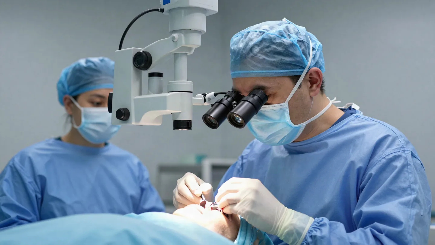
For most of the last century, glaucoma surgery meant trabeculectomy: effective, well understood, and carrying a complication profile that made surgeons wait until medical therapy had clearly failed. Minimally invasive glaucoma surgery changed that calculation — not by replacing filtration surgery, but by creating a category of intervention safe enough to offer earlier.
The term now covers a wide and occasionally confusing range of procedures. This is a map of the landscape and the logic behind choosing between them.
What makes a procedure "MIGS"
The label is descriptive rather than regulatory, but the procedures generally share an ab interno approach through a clear corneal incision, minimal disruption of the conjunctiva, a favourable safety profile and rapid visual recovery. The trade-off is usually a more modest pressure reduction than filtration surgery achieves.
That trade-off is the whole point. A procedure that lowers pressure less but risks less can be offered to a patient whose disease does not yet justify a bleb.
The trabecular approaches
These target the conventional outflow pathway by bypassing or removing the trabecular meshwork, working with the eye's existing drainage anatomy.
Stents and bypass devices
Small implants placed across the meshwork to channel aqueous directly into Schlemm's canal. Frequently combined with cataract surgery, where the incision already exists.
Goniotomy and trabecular excision
Rather than implanting, these open or remove a segment of meshwork. No device is left behind, which matters for some patients and some health systems.
Canaloplasty
Viscodilation of Schlemm's canal to restore flow through the collector channels, sometimes combined with a goniotomy in the same sitting.
The shared limitation of every trabecular approach is episcleral venous pressure: they cannot lower intraocular pressure below it, which sets a floor on what they can achieve.
The subconjunctival approaches
These create a new drainage route to the subconjunctival space, and so are not bound by that floor. They can reach lower target pressures — and, being bleb-forming, they reintroduce the concerns that come with a bleb, including scarring and the need for fibrosis management. The revision of a failed subconjunctival device is its own surgical skill.
The suprachoroidal route
Directing aqueous into the suprachoroidal space is anatomically attractive and has had a difficult commercial history, with devices withdrawn and reintroduced. Worth understanding; worth following the evidence closely before adopting.
How the choice is actually made
- Target pressure. If the target sits below episcleral venous pressure, a trabecular procedure cannot get there on its own.
- Stage of disease. Advanced field loss rarely tolerates a modest reduction and a wait to see.
- Is cataract surgery planned? Combining changes the risk–benefit arithmetic substantially.
- Angle anatomy and visualisation. Every trabecular procedure depends on a view of the angle — and on the surgeon's gonioscopic skill.
- What happens next. Preserving the conjunctiva keeps filtration surgery available later. Spending it does not.
MIGS did not make filtration surgery obsolete. It made the decision to perform it a considered one rather than an inevitable one.
Learning the angle
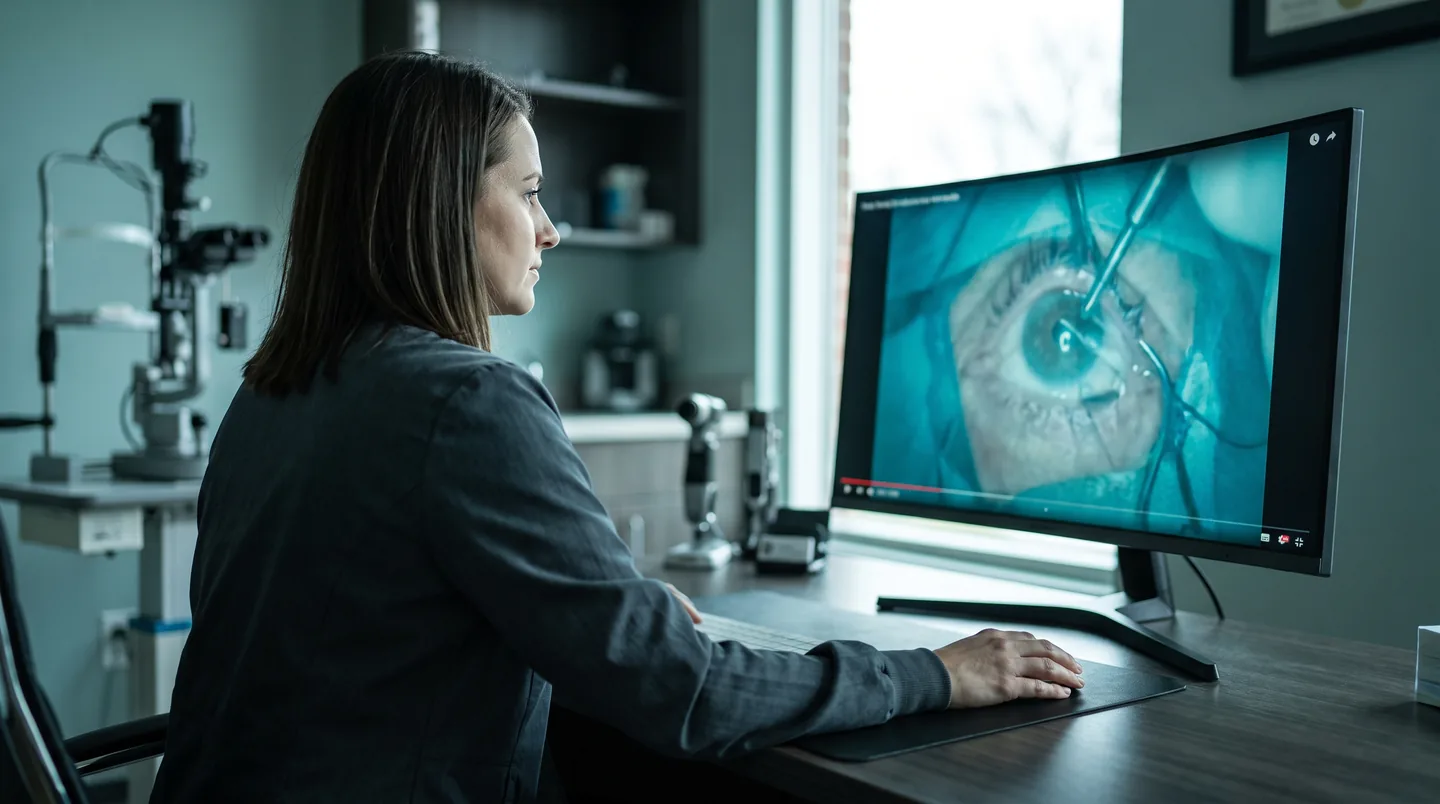
The rate-limiting skill is not the device — it is intraoperative gonioscopy. Surgeons adopting these procedures consistently report that head and microscope positioning, and reliably identifying the meshwork, take longer to master than the implantation itself. That is a skill best learned from video and wet lab before theatre.
Glaucoma courses on Ophthalmology Radar
Surgical video and structured education on filtration surgery, drainage devices, MIGS and bleb management.
In short
MIGS is a category, not a procedure, and the interesting question is never "which device is best" but "what pressure does this eye need, and what am I prepared to spend to get there". Answer that first and the device usually chooses itself.

We connect ophthalmologists worldwide to income, education, events and each other. Written to help you get more from your expertise.